
Figure 7.
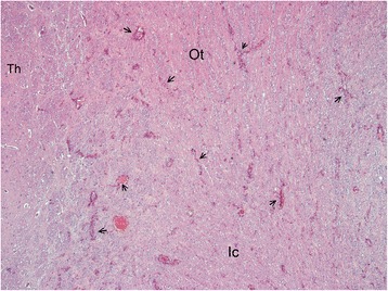
Overview histological image in the forebrain at the level of the Thalamus (Th). Diffuse pale staining and vacuolation of the white matter is seen in the internal capsule (Ic) and Optic tract (Ot). The walls of many blood vessels (arrows) in the white matter appear thickened. HE, 10X.