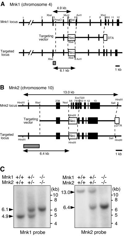
FIG. 1.
Targeted disruption of the mouse Mnk1 and Mnk2 genes. (A and B) Schematic illustration of the exon organization of the Mnk1 (A) and Mnk2 (B) genes and targeting strategy. The exons of each gene are indicated by filled boxes. The targeting vectors were designed to replace exons 5 and 6 of the Mnk1 gene or exons 5 to 9 of the Mnk2 gene with a Neor cassette. The diphtheria toxin A fragment gene (DTA cassette) used for negative selection was placed at the end of the 3′ homologous arm. Probes used for Southern blot analysis are indicated by hatched boxes. (C) Southern blot analysis of genomic DNA from WT, Mnk1+/− Mnk2+/−, and Mnk1−/− Mnk2−/− mice. DNA was digested with XbaI (left panel) or HindIII (right panel) and analyzed by Southern hybridization with Mnk1 and Mnk2 probes, respectively. As indicated in panel A, WT and targeted alleles of the Mnk1 gene were predicted to result in bands at 4.9 and 6.1 kb, respectively, whereas those of the Mnk2 gene were expected to result in 13- and 6.4-kb bands, respectively.

