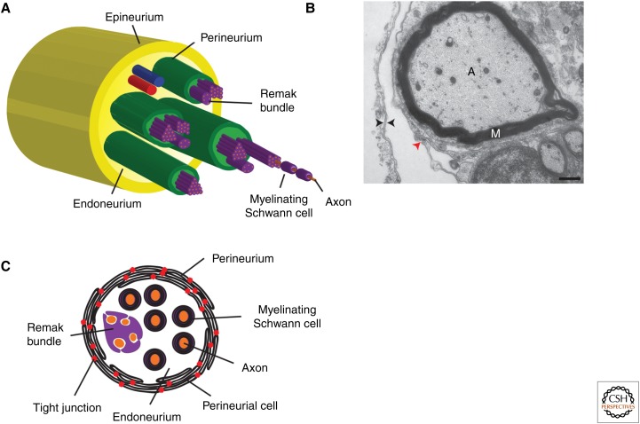
Figure 1.
Structure of peripheral nerves. (A) Diagram of the five major cell types that are found in peripheral nerves. Individual axons (orange) are ensheathed by either myelinating or Remak Schwann cells (purple). Multiple axon–Schwann cell complexes reside with the endoneurium (light green), and are ensheathed into fascicles by the perineurium (green). All fascicles within a nerve are then ensheathed in a connective tissue known as the epineurium (dark yellow). (B) Electron micrograph of the edge of a motor nerve showing that perineurial cells have a double basal lamina (black arrowheads) and are connected via tight junctions (red arrowhead). (C) Detailed schematic of an individual nerve fascicle. Multiple axon–Schwann cell complexes are ensheathed in concentric rings of perineurial glia that are connected via tight junctions (red dots). Scale bar, 500 nm. A, axon; M, myelin.

