Figure 2.
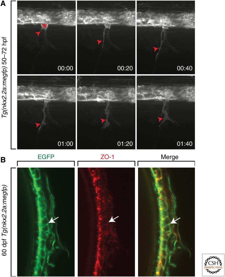
The motor nerve perineurium is composed of CNS-derived perineurial glia. (A) Frames captured from a 22-h time-lapse sequence of a Tg(nkx2.2a:megfp) zebrafish embryo beginning at 50 h postfertilization (hpf). Numbers in the lower right corners show the time elapsed from the first frame. At 50 hpf (00:00), a GFP+ perineurial process (red arrowhead) exits the CNS and is subsequently followed by a cell body (red asterisk). (B) Transverse section of a 60-d postfertilization (dpf) Tg(nkx2.2a:megfp) adult. Antibody labeling to ZO-1 shows GFP+ perineurial membranes colabeled with this tight junction marker (arrows).

