FIG. 3.
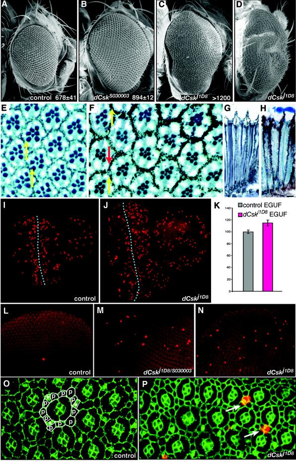
dCsk mutants show an increased proliferation. (A to D) SEMs of EGUF eye clones FRT82B Ubi-GFP control (A), dCskS030003 (B), and dCskj1D8 (C) and rear view of severely overgrown dCskj1D8 EGUF clone (D). Inset values show the average number of ommatidia per eye per genotype (n = 3 or more). (E) Adult retinal sections of control EGUF eyes. The normal complement of photoreceptor neurons can be confirmed by the 7 rhabdomeres within each ommatidium. Note that each rhabdomere array forms a trapezoid that points upward (yellow arrows). (F) Sections of adult dCskj1D8 EGUF eye clones show normal ommatidia. However, some ommatidia have reversed planar polarity, as assessed by rhabdomere assays (red arrow). (G and H) Longitudinal sections of an adult EGUF control retina (G) and a dCskj1D8 retina (H) show that dCskj1D8 ommatidia are normal in depth as well as width. Lenses were lost from some ommatidia during sectioning. (I and J) Larval eye-antennal imaginal discs from EGUF clones stained with anti-phospho-histone to highlight mitotic nuclei. The anterior is shown toward the right. For each,the eye disc is to the left and the rounded portion of tissue to the right is the antennal disc. The dotted line marks the position of the morphogenetic furrow. The very large phospho-histone-positive nuclei posterior to the furrow are within the overlying peripodial membrane and not located in the eye disc proper. Most phospho-histone-positive nuclei posterior to the furrow are within the eye disc and are part of the second mitotic wave. Note the increased size of the dCsk eye disc and the increased number of phosphohistone-positive nuclei anterior to the furrow in panel J. (K) Analysis of mitosis anterior to the morphogenetic furrow in eye imaginal discs. The number of mitotic nuclei anterior to the furrow was quantified, and results were controlled for tissue mass (see Materials and Methods). Results are normalized to those of control EGUF tissue. Bars represent standard errors. (L to N) Pupal eye discs at 26 h after puparium formation stained with anti-phospho-histone (bright red) to highlight mitotic nuclei. All nuclei stain faintly (background red) with the antiphosphohistone antibody. (O and P) Pupal eye discs at 26 h after puparium formation stained with anti-phospho-histone antibody (red) to mark mitotic cells and anti-armadillo antibody (green) to mark cell boundaries. Each individual ommatidium is composed of 8 photoreceptors (located basally out of the plane of focus), 4 overlying cone cells, and 2 primary pigment cells. (O) Cells that make up the interommatidial lattice surrounding a single unit ommatidium are outlined in white; lattice cells include bristle groups (*) and secondary and tertiary pigment cells (p). (P) The excess mitotic cells in dCskj1D8 tissue are located within the interommatidial lattice (arrows) as determined by nuclear position and cell positions.

