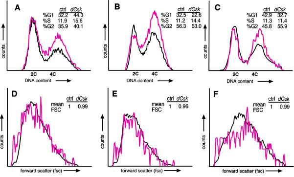
FIG. 4.
Flow cytometry analysis shows a cell-autonomous proliferative defect in dCsk mutant cells. (A and B) Flow cytometry analysis on wild-type (black) and dCskj1D8/S030003 mutants (pink). The DNA content of eye-antennal disc cells (A) and wing disk cells (B) from 126- to 128-h-AED larvae is shown. We observed similar differences between wild-type and dCsk mutant cell cycle profiles in more than seven repeat experiments. (C) DNA content of dCskj1D8 mutant cells from mitotic clones and neighboring wild-type and heterozygous cells from eye-antennal tissue (120 h AED). Forward scatter (FSC) profiles of eye-antennal cells from panel C (D) and separate analysis of cells with 2C (E) and 4C (F) DNA content. FSC is a relative measurement of cell size (51). The mean FSC of dCsk mutant cells in relation that of to control (ctrl) cells is indicated.

