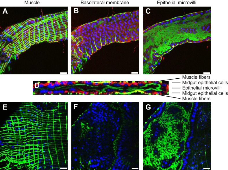
Fig. 6.
Immunostaining of dissected A. aegypti anterior and posterior midgut. Confocal slices of the muscle, basolateral membrane and epithelial microvilli of the anterior (A–C) and posterior (E–G) midgut. (D) Confocal stack through the thickness of the anterior midgut. Red: GluCl; green: actin; blue: DAPI. Scale bars: 50 µm.

