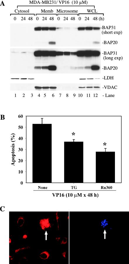
FIG. 8.
Potential mitochondrion-ER cross talk in VP16-induced apoptosis. (A) BAP31 was preferentially cleaved in the population of ER that cofractionated with the mitochondria. Vehicle-treated or VP16-treated MDA-MB231 cells were used to obtain various fractions indicated. Equal amounts of proteins (40 μg/lane) were used in Western blotting for the molecules indicated. Two different exposures (i.e., short and long exp.) for BAP31 were shown. Note that only VDAC-OMM was shown for the sake of simplicity. Memb, membrane; WCL, whole-cell lysate. (B) Depletion of ER Ca2+ store or inhibition of mitochondrion uptake of Ca2+ inhibited VP16-induced apoptosis. MDA-MB231 cells were pretreated with either vehicle (ethanol) or 50 nM TG or 50 μM Ru360 for 1 h. After removing these chemicals, cells were treated with VP16 (10 μM) for 48 h. At the end of treatment, cells were scored for apoptosis by DAPI staining. On average, 250 to 400 cells were counted for each condition and data represent means ± standard deviations. *, P < 0.01 (Student's t test). (C) Mitochondrial fragmentation during VP16-induced apoptosis. MDA-MB231 cells treated with VP16 (10 μM for 48 h) were labeled live with Mitotracker and DAPI. The arrows indicate DAPI-positive (i.e., apoptotic) cells that show fragmentation of the mitochondria. The micrographs shown are representative of ∼400 hundred images analyzed.

