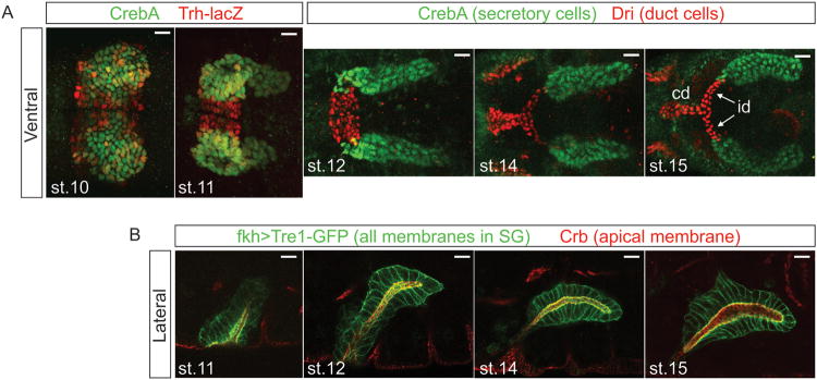
Figure 1.

Confocal images of the embryonic SG. (A) Ventral views of the SG stained with nuclear markers. The secretory portion of the SG forms from two placodes of cells on the surface of the embryo, with the duct precursors located between the two secretory placodes (st 10). At this stage, the expression of duct (red) versus secretory (green) markers is not so clear. During st 11, the gland invaginates into the embryo. At this stage, the distinction between duct (red) and secretory markers (green) is more evident. As development progresses (st 12-15), the secretory tubes elongate, and the individual duct (id) and common ducts (cd) form. (B) Lateral views of SG during elongation and migration. All membranes are marked in green, with the apical membrane specifically marked in red. Following invagination, the SG moves dorsally (st 11) and then turns (st 12) and migrates posteriorly. Posterior migration continues (st 14-16) until the SG reaches its final resting place. Throughout this dynamic process, the SG migrates as an intact fully polarized tissue.
