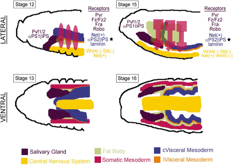
Figure 5.
The SG contacts or comes close to several tissues as it migrates to its correct final position in the embryo, including the circular (c) visceral mesoderm (cVM), the longitudinal (l) visceral mesoderm (lVM), the fat body, the somatic musculature and the central nervous system. The cVM provides a suitable substrate for posterior SG migration through the expression of αPS2βPS integrin that binds a secreted laminin also expressed in the cVM. The SG expresses αPS1βPS integrin, which also binds the secreted laminin. Both the integrins and laminin are essential for posterior migration (starred). The lVM migrates between the SG and the cVM to detach these two cell types. The SG also expresses several receptor genes, which allow it to properly navigate to its final correct position in response to local sources of the corresponding ligands. In turn, the SG is likely to also provide cues for the migration of other cell types in the embryo. For example, the fat body migrates over specific parts of the SG at late embryonic stages.

