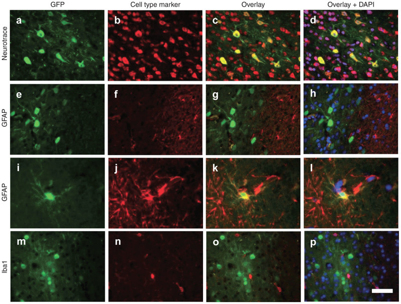
Figure 3.
Predominantly neuronal transduction of intrathecal AAV9. (a,e,i,m) Cortical brain sections from animals treated by intracisternal injection were imaged for native GFP fluorescence. Images are overlaid with a (b–d) fluorescent Nissl stain (NeuroTrace) for detection of neurons, (f–h,j–l) GFAP immunostaining for astrocytes, or (n–p) Iba1 staining for microglia. (d,h,l,p) Counterstain is DAPI. Scale bar = 50 µm.

