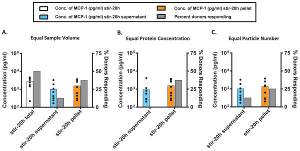
Figure 2.
Peripheral blood mononuclear cells (PBMC) from 8 human donors were incubated with solutions of mAb2 particles (see Figure 1) and examined for the release of MCP-1. MCP-1 was measured by electro-chemiluminescence at the early phase (20 h incubation) at (A) equal sample volume, (B) equal protein concentration, and (C) equal particle numbers. The average concentration (n=8, colored bars) of MCP-1 and percentage of donors that responded (gray bars) to the aggregated mAb (two fold above the unstressed mAb2) is shown. The black dots represent the concentration of MCP-1 secreted by each individual donor. Different aggregate samples are shown horizontally as follows; stir-20h total (white), stir-20h supernatant (enriched in nanometer size particles; light blue), and stir-20h pellet (enriched in micron sized particles; orange); see Figure 1. The media and buffer-stressed controls responded far below the threshold, and the LPS positive control responded much more intensely (SI≫2.0) than the aggregated protein.

