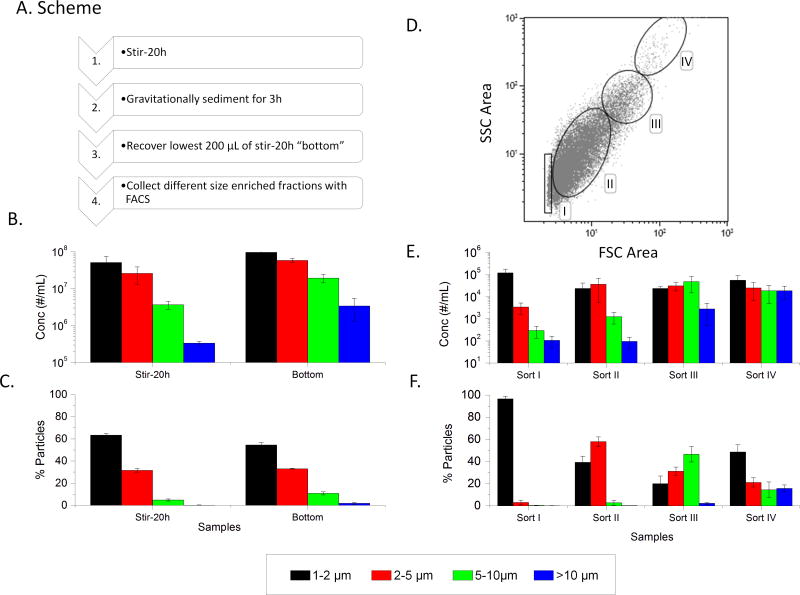
Figure 3.
Enrichment of different micron sized mAb2 particle populations using FACS separation. (A) Flowchart of experimental steps. (B) Subvisible particle counts in stirred sample (stir-20h) and bottom fraction after gravitational settling (bottom sample) as measured by MFI. (C) The percentage of particles in each size bin for these same two samples. (D) Upon FACS sorting the bottom sample, a two dimensional dot plot of response from forward vs side scattering signals (FSC Area vs. SSC Area) is generated with gatings labeled Sort I–IV, (E) Sorts I–IV were analyzed for subvisible particle distribution with MFI. (F) The percentage of particles in each size bin were determined. The graphs represent the average of three separate experiments (N=3) with the error bars representing one standard deviation.

