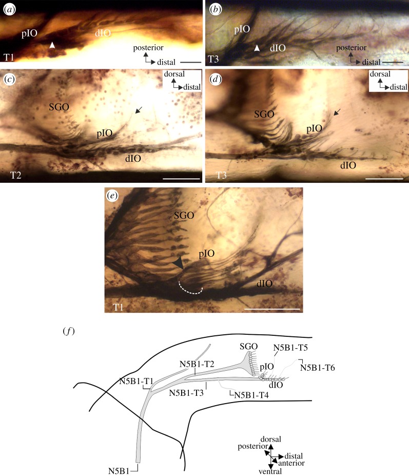
Figure 2.
Neuroanatomy of the IO in the legs of T. neglectus. (a,b) Neuron somata of the sensilla in the dIO are arranged in series in a foreleg. The most proximal soma in the dIO is indicated by arrowhead. (c,d) Neurons of the pIO are positioned proximally and dorsally to the dIO. Dendrites of the pIO point dorsally with their distal segments (arrow). Neurons of the dIO extend distally into the tibia. (e) A section of the pIO may be supplied by a small, common nerve branch (arrowhead), as shown here from a foreleg preparation. Note that only the dorsal pIO neurons are supplied by this branch, the other pIO neurons (dotted semicircle) form no distinct nerve branch. (f) Drawing reconstruction of the innervation of sensory organs by nerve 5B1, showing the consensus branching pattern of the sensory nerve N5B1 in the legs of T. neglectus. Axes are given for the tibia. All preparations viewed from anterior. Scale bars: (a) 200 μm, (b,c) 100 μm, (e,f) 50 μm.

