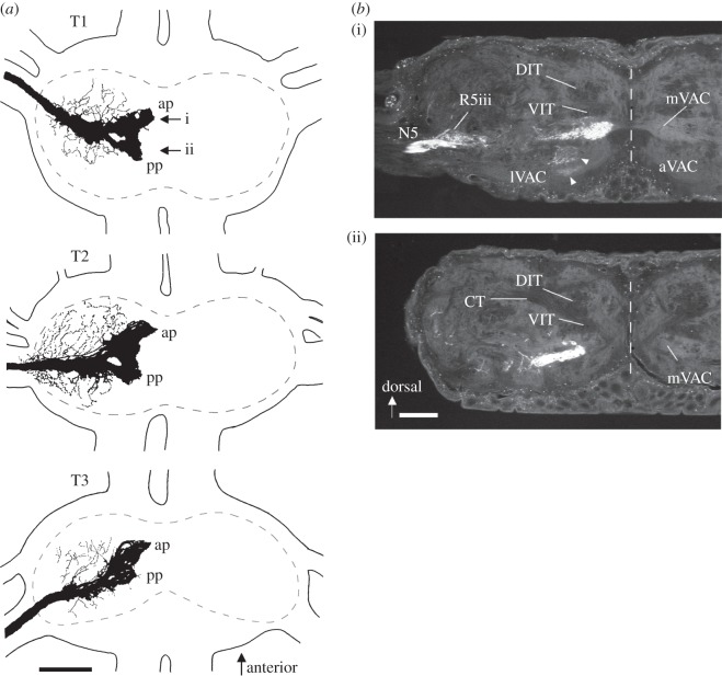
Figure 4.
Central projection of the nerve 5B1. (a) Wholemount drawings of preparations from the three thoracic ganglia. The sensory afferents terminate in a dense projection close to the midline, which has an anterior projection (ap) and a posterior projection (pp). The neuropile outline is hatched. Scale bar, 200 μm. (b) Histological sections of a prothoracic ganglion (with the nerve 5B1 filled with Lucifer Yellow), at the level of the anterior (i) and posterior (ii) projection indicated by arrows in (a). Arrowheads indicate terminations in the aVAC, which are typical for sensory hairs; these projections are entirely separated from that of the SGO complex and are not included in (a). Scale bar, 100 μm. Midline is indicated by hatched line. T1–T3, prothoracic, mesothoracic and metathoracic ganglion, respectively; ap, anterior projections; pp, posterior projections; DIT, dorsal intermediate tract; VIT, ventral intermediate tract; CT, C-tract; N5, nerve 5; R5iii, third root of the N5; aVAC/lVAC/mVAC, anterior/lateral/median ventral association centre.

