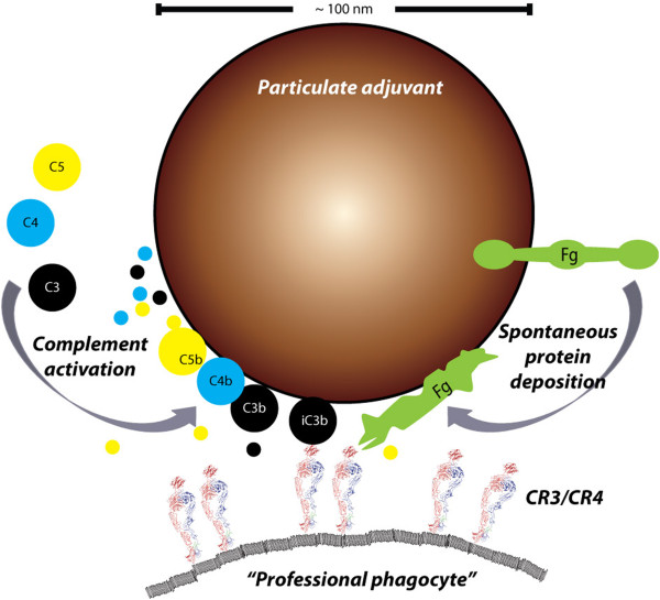
Figure 2.

The sources and consequences of plasma protein deposition on particulate adjuvants. Deposition of complement is well-described for liposomes and is likely to happen on the surface lipid adjuvants as well [142]. The deposition of complement on aluminum oxide particles is less characterized [148]. As clear from the schematic, a particle with a diameter of ~100 nm presents certain space and topological restraints, which may influence both the complement activation and actually capacity for carrying deposited protein as detailed elsewhere [134]. Following activation of the complement components C3, C4, and C5 these are covalently bound to target surfaces concomitantly with the proteolytic release of small peptides from the molecules (indicated with colors). This release depends on the pathway of complement activation and may hence create a signature for adjuvants. In this scenario, which does not explicitely involve the vaccine antigen, the adjuvants act as by-standers creating a milieu of immunostimulatory peptides. Proteolytic processing of the C3b fragment creates the iC3b fragments, which is ligand for both CR3 and CR4 and may thus support phagocytosis by “professional phagocytes” [161] through the connection of these receptors with cytoskeleton. In addition to complement, surface-adsorbed fibrinogen is a ligand for CR3 and CR4, probably in part because of the structural denaturation of the protein as a consequence of the surface adsorption, which enhances the interaction with these receptors [155, 156]. Sizes are indicated approximately to scale based on data presented in Refs. [134, 166, 167].
