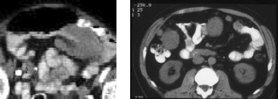
Figure 8.

Embolic metastases. (a)Post contrast axial CT scan in a 60-year-old male with malignant melanoma showing a well defined embolic serosal deposits causing small bowel obstruction. (b) Axial CT scan in a 40-year-old male with leiomyosarcoma of the retroperitoneum showing multiple well defined soft tissue masses adjacent to the bowel in the mesenteric fat.
