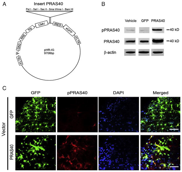
Fig. 1.
Construction of lentiviral vectors of PRAS40 and its expression in primary neuronal cultures. A. Schematic backbone of lentiviral vector of pHR’tripCMV–IRES–eGFP. B. Western blot confirmation of the effects of gene transfer in primary neuronal cultures on protein expression of p-PRAS40 and PRAS40. Protein levels of both PRAS40 and p-PRAS40 were increased in cells transfected with PRAS40. All protein bands were cut from the same gel (see Supplementary Fig. 1 for the original gel). C. Triple staining of GFP, PRAS40 and DAPI in a mixed primary neuronal culture transfected with PRAS40 confirms the successful transfection of neurons. Scale bar, 50 μm.

