
Figure 1.
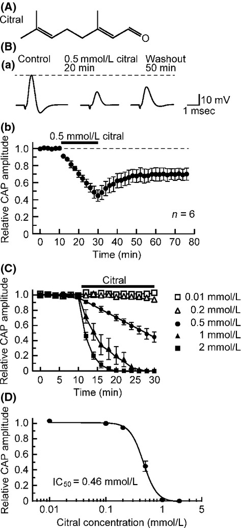
Effect of citral on compound action potentials (CAPs) recorded from frog sciatic nerve fibers. (A) The chemical structure of citral. (B) Citral at a concentration of 0.5 mmol/L reduces CAP peak amplitudes. (a) Recordings of CAPs in the control, at 20 min after exposure to citral, and thereafter 50 min in the absence of citral. (b) Average time course of changes in CAP peak amplitudes following exposure to citral for 20 min, relative to control. In this and subsequent figures, each point with vertical bars represents the mean and SEM, and the dotted line denotes the control value. (C) Average time courses of CAP peak amplitude reductions produced by citral at 0.01–2 mmol/L; data at each concentration were obtained from 3 to 4 sciatic nerves. Solid lines in this graph were arbitrarily drawn. (D) The peak amplitudes of CAPs recorded from sciatic nerve fibers treated with citral at various concentrations for 20 min, relative to control, which were plotted against citral concentration. Each of the data points was obtained from 3 to 4 sciatic nerves. This concentration–response curve was drawn according to the Hill equation (IC50 = 0.46 mmol/L; nH = 3.6).