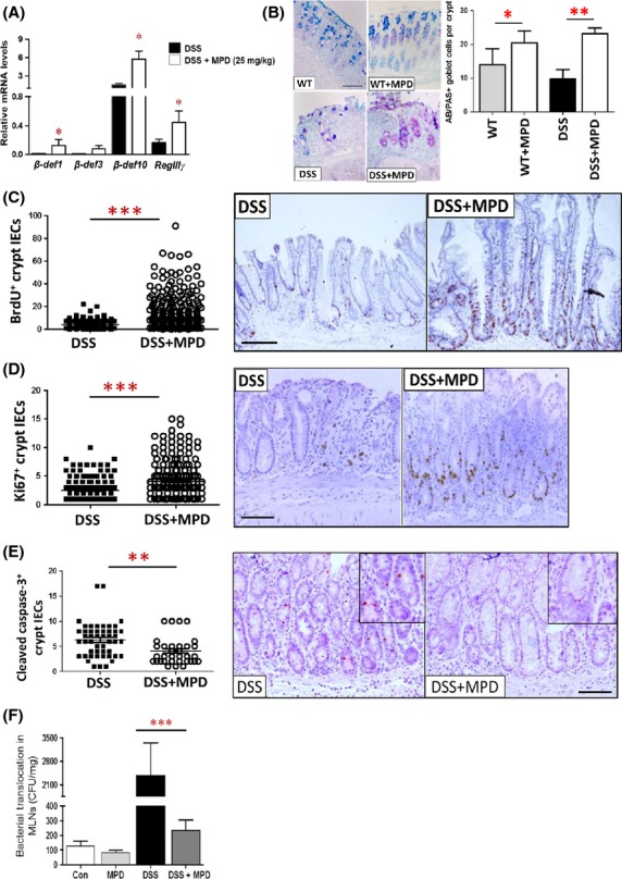
Figure 4.

MPD administration promotes antimicrobial peptide expression and increases intestinal epithelial proliferation to enhance intestinal barrier function. Intestinal inflammation was induced by 2.5% DSS for 7 days followed by 5 days of water recovery. MPD was given to mice at 25 mg/kg daily on day 3 together with DSS for 10 days. (A) Colonic mucosal RNA was then extracted; the levels of AMP mRNA were quantitated by qPCR. *P < 0.01. (B) Goblet cells were stained with AB/PAS staining, and numbers of goblet cells were counted and expressed as AB/PAS+ cells per crypts. (C–E) IEC proliferation or apoptosis was determined with BrdU labeling (C) and Ki67 immunohistochemistry (D), or Cleaved Caspase-3 immunohistochemistry (E). Numbers of proliferative IEC or apoptotic IEC was counted in the well-orientated crypts. Results are expressed as mean ± SEM (n = 5 − 6). *P < 0.05, **P < 0.01 versus controls. (F) Intestinal inflammation was induced by 2.5% DSS for 7 days followed by 5 days of water. MPD was given to mice at 25 mg/kg daily on day 3 together with DSS for 10 days. Bacterial translocation to MLN was measured and expressed as colony-forming unit per mg lymphatic tissue. Results are expressed as mean ± SEM, n = 6 per group. ***P < 0.001 versus DSS colitis group, bar = 200 um.
