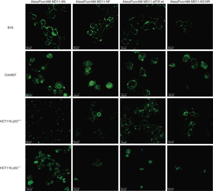
Figure 3.
Confocal laser scanning microscopy images of living B16 (0.5 hour), Colo857 (1 hour), HCT116 p53+/+, (2 hours) and HCT116 p53−/− (2 hours) cells incubated with AlexaFluor 488-labeled MD11-AN, MD11-NF, MD11-wt eIF3f, and MD11-K510R fusion protein (10 nmol/l). 16 successive optical slices were captured along the cellular z-axis with a step of 1 µm. The presented images correspond to the middle plan of cellular z-axis sectioning.

