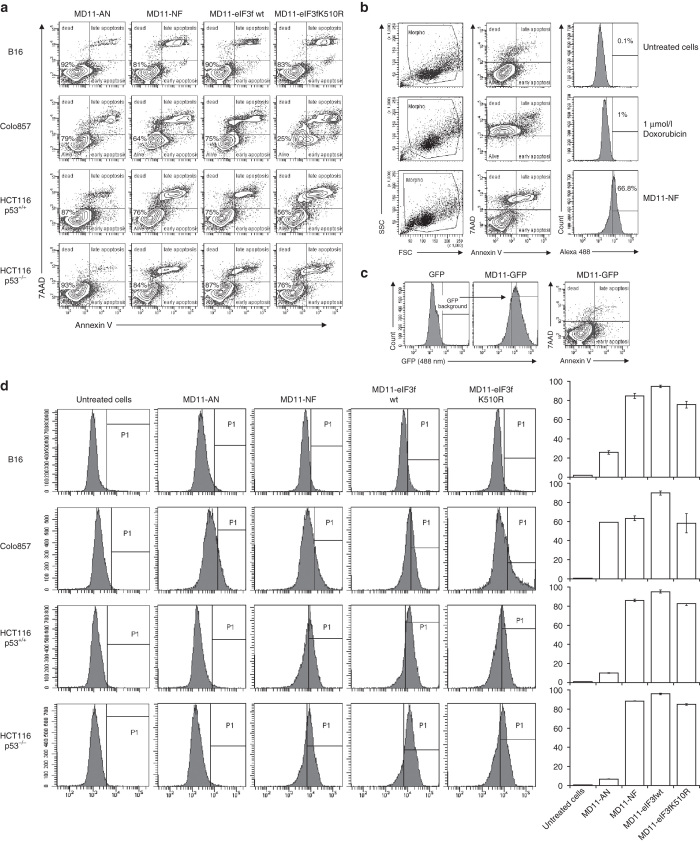
Figure 4.
Annexin V-7AAD apoptosis assay and protein delivery efficiency analysis by flow cytometry. (a) B16, Colo857, HCT116 p53+/+, and HCT116 p53−/− cells were incubated with AlexaFluor 488 labeled proteins (10 nmol/l) and stained with Annexin V-APC and 7-AAD. Alive cells show negative staining for Annexin V and 7-AAD. Cells undergoing apoptosis show positive staining for Annexin V and negative for 7-AAD, while cells positive for both Annexin V and 7-AAD are either in the end stage of apoptosis, undergoing necrosis or already dead. (b) Examples of FACS profiles of untreated cells and doxorubicin-treated cells, used as controls for viability/toxicity setting gates. (c) Representative plot of HCT116 p53−/− cells incubated with 0.3 µmol/l MD11-eGFP, showing no toxic effect associated to the MD11-mediated protein internalization. (d) Evaluation of delivery efficiency of AlexaFluor 488-labeled recombinant proteins in selected cells. Histograms represent percentages of internalized MD11-AN, MD11-NF, MD11-wt, and MD11-K510R proteins in target cells.

