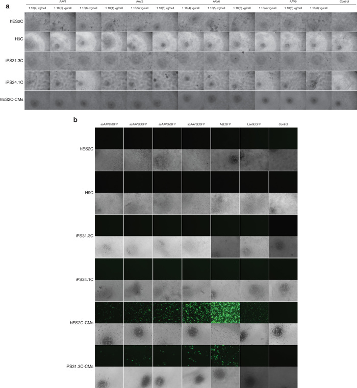
Figure 2.
Representative images of different pluripotent (hES2, H7, hiPS31.3, hiPS24.1) and differentiated (hES2-CM and hiPS31.3-CM) cells transduced with adeno-associated virus (AAV), adenoviral, and lentiviral vectors. (a) Representative images of hES2, H7, hiPS31.3, hiPS24.1 cells, hES2-CM, and hiPS31.3-CM infected with ssAAV vectors carrying the luciferase gene. ssAAV serotypes 2 and 6, and highest concentrations of serotypes 1 and 9 induced cell death in hES2 and iPS31.3 and iPS24.1 cells. AAVs did not appear to affect the viability of differentiated cells. (b) Representative images of hES2, H7, hiPS31.3, hiPS24.1 cells, hES2C-CMs, and hiPS31.3C-CMs infected with scAAVs 2 and 6, adenoviral and lentiviral vectors carrying the GFP gene. Images of the infections using the highest concentration of virus per cell are shown. High levels of AAVs reduced the survival of hES2 and hiPS31.3 cells. No cell death was observed in hES2C- and hiPS31.3C-derived cardiomyocytes, which also exhibited high levels of transduction compared to their undifferentiated parental cell lines.

