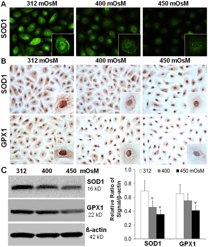
Fig 5. The reduced production of anti-oxidative enzymes SOD1 and GPX1 in HCECs exposed to hyperosmotic stress.
Primary HCECs exposed to media at 312, 400 and 450 mOsM for 24 hours were performed for immunofluorescent staining (A), or immunohistochemical staining (B), or lysed in RIPA buffer for Western blotting (C), to determine the protein production of SOD1 and GPX1. Data showing mean±SD, n = 4, V P<0.05, VV P<0.01, compared with 312 mOsM normal control.

