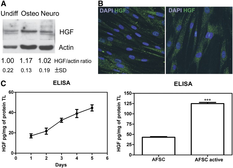
Figure 1.
HGF expression by human AFSCs (hAFSCs). (A): Western blot analysis with anti-HGF revealed total lysates of undifferentiated hAFSCs, after 2 weeks in culture with osteogenic medium, and after 3 weeks in culture with neurogenic medium. Actin detection was performed to show the amount of protein loaded in each lane. Presented data are representative of three independent experiments, and the gray density values were normalized to that of actin. (B): Representative immunofluorescence images obtained at different magnifications of undifferentiated hAFSCs labeled with DAPI (blue) and HGF (green). Scale bar = 10 μm. (C): ELISA quantification (normalized to protein content) of HGF in media obtained from hAFSCs after 1–5 days in culture (left) and compared with hAFSCs preactivated for 24 hours and after 5 days in culture (right). Student’s t test showed a significant difference between samples. ∗∗∗, p ≤ .0001. Presented data are the average of three independent experiments. Abbreviations: AFSC, amniotic fluid stem cells; DAPI, 4′,6-diamidino-2-phenylindole; ELISA, enzyme-linked immunosorbent assay; HGF, hepatocyte growth factor; Neuro, neurogenic; Osteo, osteogenic; TL, total lysate; Undiff, undifferentiated.

