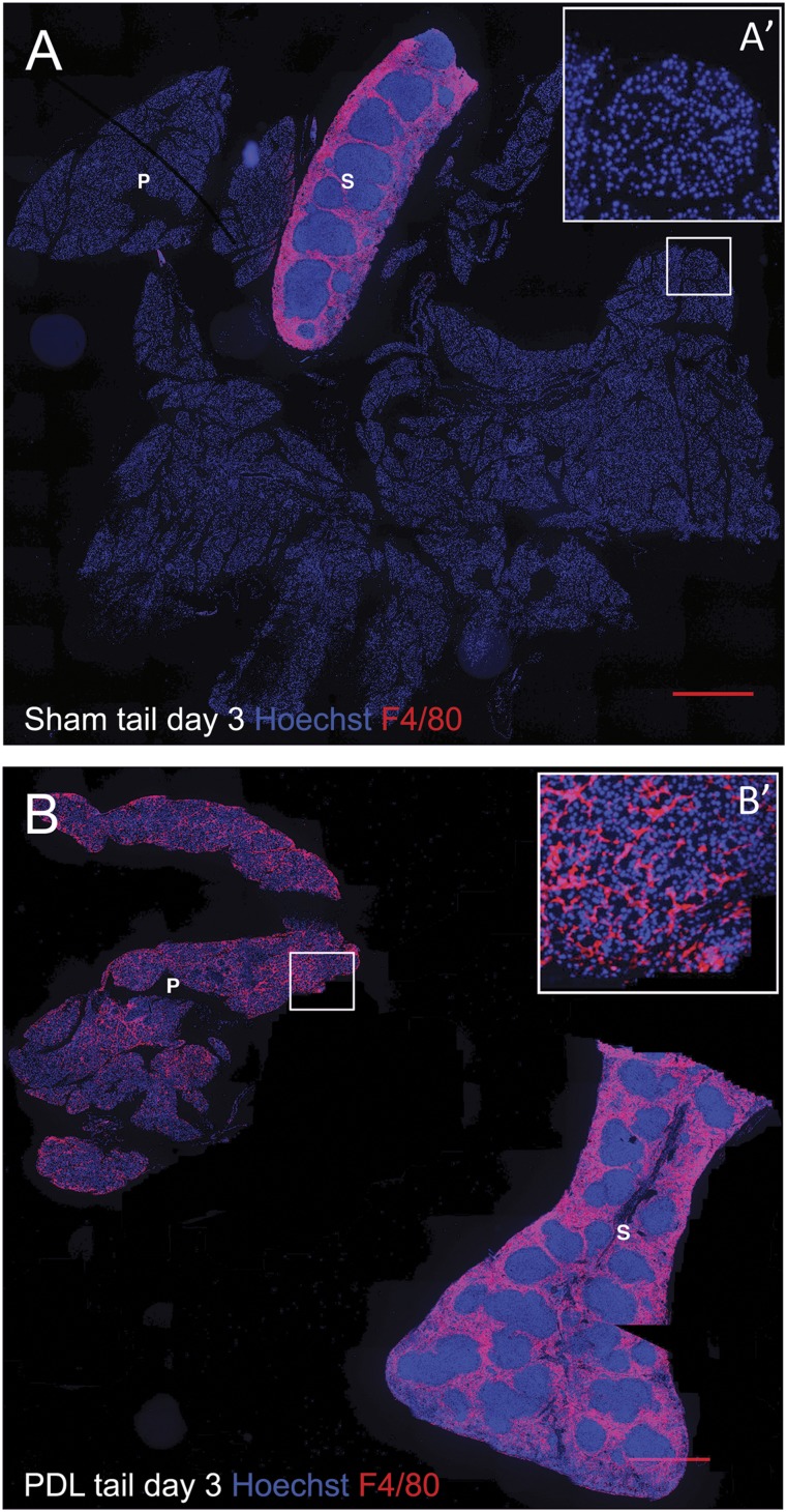
Figure 4.
Massive macrophage infiltration in the pancreas after partial pancreatic duct ligation. Pancreas tail and spleen sections from day 3 sham-operated (A) and PDL (B) mice, stained for nuclei (Hoechst, blue) and F4/80 (red). (A′, B′): Higher magnification of the area depicted by the squares in (A) and (B). Scale bars = 500 μm. Notably, PDL results in acinar cell loss and thus a decrease in the total pancreas tail area versus sham-operated mice. Abbreviations: P, pancreas tail; PDL, pancreatic duct ligation; S, spleen.

