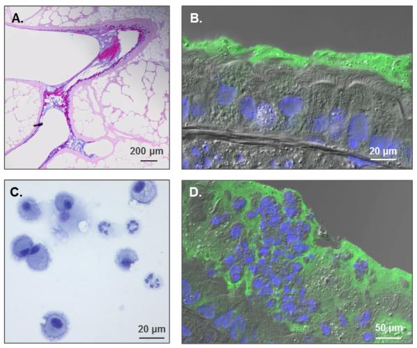
Fig. 1. Formation of mucus plaques and neutrophilic inflammation in the • ENaC mouse model.
(A.) AB-PAS stain of βENaC lungs revealing mucus plugs at airway branching. (B.) Immunohistochemistry (IHC) displaying PCL collapse and mucin accumulation on airway surfaces (green=Muc5b, Blue=DAPI). (C.) Inflammatory cells from a βENaC mouse bronchoalveolar lavage showing a mixture of macrophages and neutrophils. (D.) IHC exhibiting inflammatory cells trapped in mucus.

