
Fig. 1. Schematic representation of MARTX toxins expressed by V. cholerae strains generated for this study.
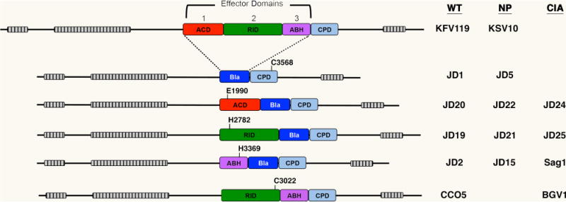
Domain organization of each toxin is shown to scale with grey-hatched bars representing the locations of the MARTX repeat regions. Effector domains are: the actin cross-linking domain (ACD, red), the Rho-inactivation domain (RID, green), and the alpha-beta hydrolase domain (ABH, purple). The location of surrogate effector domains beta-lactamase (Bla without secretion signal) is shown in blue. The cysteine protease domain (CPD) required for autoprocessing to release effector domains and/or Bla to eukaryotic cells after translocation is depicted in light blue. The relative position of catalytic residues for each effector domain and the CPD are shown numbered according to the full-length 4545 aa RtxA protein as annotated by Lin et al. (1999) and verified by Dolores & Satchell (2013). Key at right indicates V. cholerae strain designations for rtxA arrangements in isogenic ΔhapAΔhlyA background that is defined as wild type (WT) in this study. Those that carry the C3568A mutation in CPD for non-processing (NP) or catalytically inactivating (CIA) mutations as indicated in the diagram. Detailed genetics of strains are listed in Table 1.