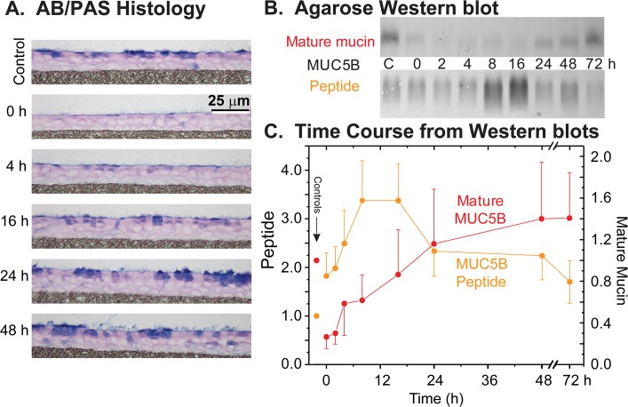
Fig 2. Recovery of mucin stores in HBE cell cultures following agonist (ATPγS, 100 μM) induced discharge.
A. HBECCs examined by AB/PAS histology. HBECCs were fixed before (control) and periodically after exposure to agonist, and mucin stores were revealed by AB/PAS staining. B. Sample agarose Western blots. Whole cell extracts of HBECCs sampled at the indicated times were subjected to electrophoresis in agarose gels, vacuum blotted, and the blots probed for MUC5B with domain-specific antibodies. ‘Immature’, non-glycosylated mucin peptides were detected with an antibody to the PTS/mucin repeat domains, and ‘mature’, fully glycosylated mucins with antibodies to the Cys-rich domains within the glycosylated mucin domains. C. Time course of MUC5B peptides and mature mucins. Mucin quantitation was achieved by densitometry of agarose Western blots as in Panel B; all results are expressed relative to their respective controls. Left axis = MUC5B peptide as the immature, non-glycosylated form of the mucin, right axis = mature MUC5B glycoprotein. Time 0 = 45 min post-ATPγS, after cultures were washed to remove the secreted mucus; Controls (‘C’ in Panel B) = non-treated HBE cells. Data expressed as the mean ± SE (n = 7–9 sets).

