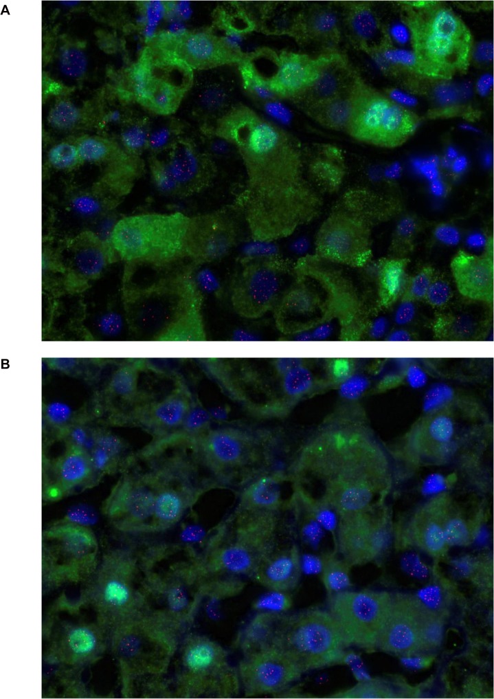
Fig 6. (a) Nuclear and cytoplasmic HBcAg staining detected by Q-FISH in liver from a representative patient with chronic HBV infection.
HBcAg stains bright green, nuclei stain blue with DAPI and telomeres stain pink. (b) Nuclear HBcAg staining detected by Q-FISH in liver from a representative patient with chronic HBV infection. HBcAg stains bright green, nuclei stain blue with DAPI and telomeres stain pink.

