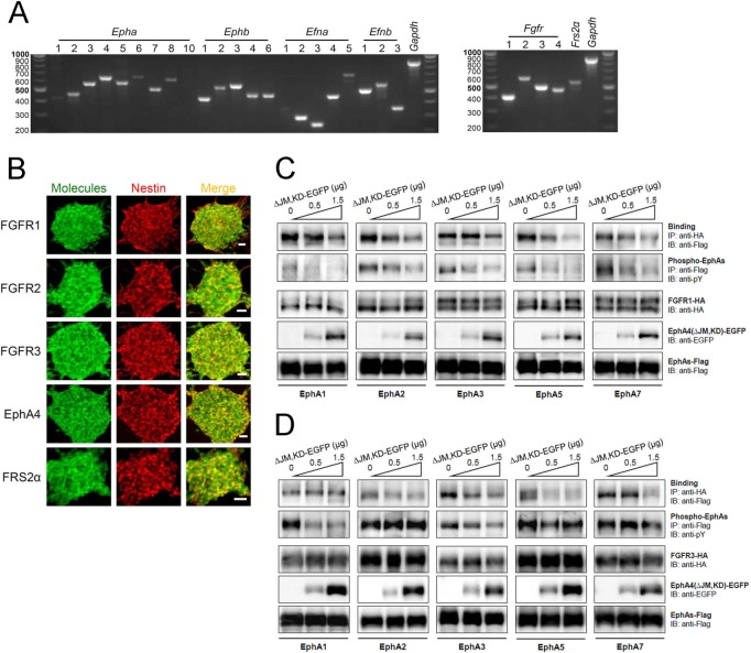
Fig 1. Expression and interaction of Ephs and FGFRs.
(A) Expression of all Eph receptors, ephrin ligands, FGFRs and related molecules in mouse embryonic NSPCs. RT-PCR was performed with equal amounts of total RNA isolated from mouse NSPCs. Fragment lengths are indicated on the left in base pairs. (B) Co-expression of FGFRs, EphA4 and FRS2α (green, left panel), respectively, with the neural stem cell marker nestin (red, middle panel) in cultured neurospheres. Merged images are shown in right panels. Neurospheres were cultured on PLL-coated plates for a short time, fixed and immunostained. Immunofluorescent images were detected using a confocal microscopy with an appropriate optical filter. (C, D) Inhibition of FGFR-EphA binding with a dominant-negative EphA4 molecule, EphA4(ΔJM,KD), tagged with enhanced green fluorescence protein (ΔJM,KD-EGFP). FGFR1-HA (C) and FGFR3-HA (D) were co-expressed with Flag-tagged EphAs (EphA1, 2, 3, 5 and 7), respectively, and increasing doses of ΔJM,KD-EGFP in HEK293T cells. Binding of FGFR-HA with EphAs-Flag was examined with immunoprecipitation (IP) followed by SDS-PAGE and immunoblotting (IB) using the antibodies shown.

