
Fig 4. ERK1/2 activation mediates the anti-melanogenic activity of hUCB-MSC-CM in melanocytes.
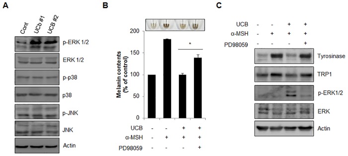
(A) Melan-a cells were incubated with hUCB-MSC-CM (UCB#1, #2) for 24 hr and analyzed protein expression by Western blotting with indicated antibodies. (B, C) Melan-a cells pre-treated with α-MSH (1 μM) were incubated with hUCB-MSC-CM in the presence or absence of PD98059 (20 μM) for 24 hr. Then, the cells were harvested (upper image) to measure the cellular melanin contents (B). The protein expressional level of tyrosinase, TRP1, p-ERK 1/2 and ERK 1/2 in the cells was further analyzed by Western blotting (C). The data were obtained from least three independent experiments and values are presented as the means ± S.E.M. (n>3, * p<0.05).