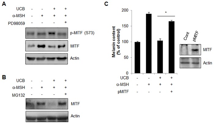
Fig 6. hUCB-MSC-CM induces proteasomal degradation of MITF mediated by ERK phosphorylation.
(A) Melan-a cells pre-treated with α-MSH (200 nM) for 24 hr were further incubated with hUCB-MSC-CM (UCB) in the presence or absence of PD98059 (20 μM) for 24 hr. Then, the expression of phosphorylated MITF at Serine 73 and total MITF protein was examined with Western blot analysis. (B) Melan-a cells pre-treated with α-MSH (200 nM) were incubated with UCB with or without MG132 (5 μM) for an additional 24 hr. Then the MITF level was examined by Western blot analysis. (C) Human MNT-1 melanoma cells transiently transfected with pEGFP (pCont) or pEGFP-MITF (pMITF) were treated with α-MSH (1 μM). After 24 hr, the cells were further incubated with UCB for additional 24 hr. Then the cells were harvested to measure the cellular melanin contents. The over-expression of MITF was confirmed by Western blotting with anti-GFP antibody. Data represent ± standard error of the mean (S.E.M.) from more than three independent experiments, n = 3,* p<0.05).

