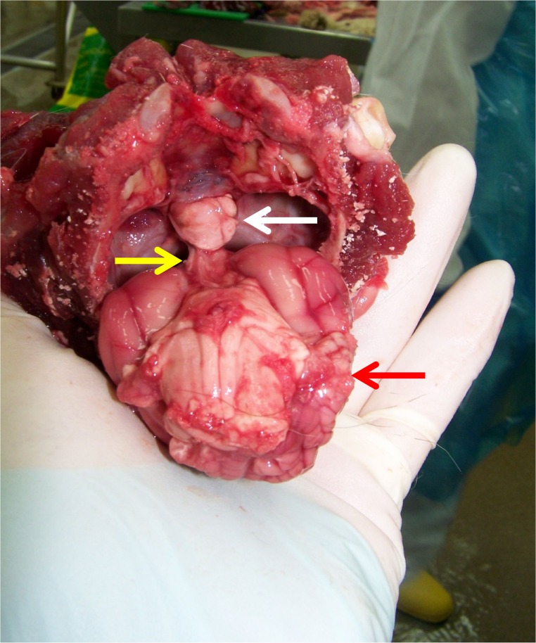
Fig 8. Macroscopic necropsy picture of the brain of a diabetic cat with confirmed HS.
This picture shows the ventral aspect of the brain of a cat with HS as it was removed from the cranial cavity and a clearly enlarged pituitary falling out of the sella turcica (cranium is held upside down; white arrow: pituitary gland; yellow arrow: pituitary stalk; red arrow: cerebrum). Histopathology confirmed the presence of an acidophilic macroadenoma.

