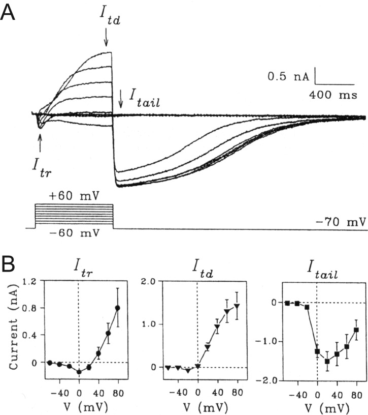
Figure 6.
Ca2+-activated Cl− currents triggered by Ca2+ entry through voltage-gated Ca2+ channels in rat pulmonary artery smooth muscle cells (PASMCs). A, Typical family of whole-cell currents (top traces) recorded from a single PASMC with the voltage-clamp protocol shown below the traces. The cell was bathed in a solution containing 1.8 mM Ca2+. B, Mean current-voltage relationships for currents measured as indicated in A and labeled as the early transient current (Itr) measured between 50 and 90 ms, the time-dependent current (Itd) measured at the end of the depolarizing step (between 750 and 790 ms), and the tail current (Itail) measured immediately after repolarization to the holding potential of −70 mV (between 900 and 940 ms). Each data point represents a mean ± SEM (n = 13). Reproduced from Yuan41 with permission from the American Physiological Society.

