Figure 8.
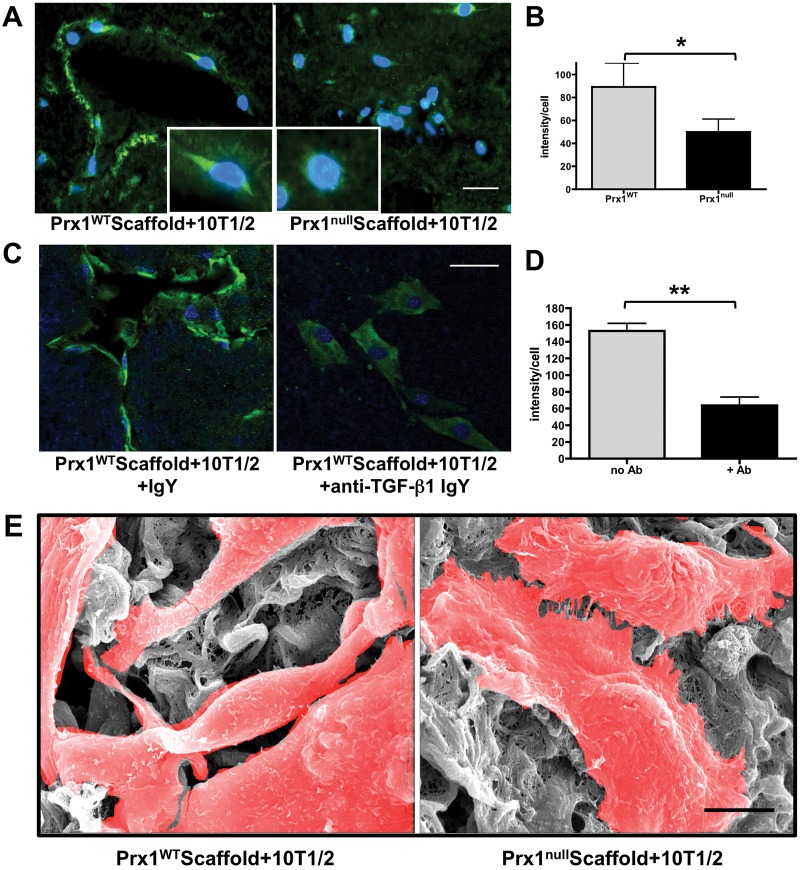
Prx1-dependent extracellular matrix regulates lung vascular smooth muscle cell differentiation. A, Immunofluorescence staining for α–smooth muscle actin (SMA; green) in 10T1/2 cells seeded within E17.5 Prx1WT (left) or Prx1null (right) lung scaffolds. Insets show enlarged 10T1/2 cell images. Nuclei stained blue by 4′,6‐diamidino‐2‐phenylindole dihydrochloride (DAPI) staining. Scale bar = 20 μm. B, Fluorescence intensity of α-SMA immunostaining on frozen sections of 10T1/2 cells cultured in Prx1WT and Prx1null scaffolds (A) was quantified using ImageJ. One asterisk indicates P < 0.05. C, Immunofluorescence staining for α-SMA (green) of 10T1/2 cells seeded in Prx1WT scaffolds, treated with either control immunoglobulin (Ig) Y (left) or transforming growth factor (TGF)–β function-blocking IgY (right). Nuclei stained blue by DAPI. Scale bar = 20 μm. D, Fluorescence intensity of α-SMA immunostaining on frozen sections of 10T1/2 cells cultured in Prx1WT scaffolds with or without function-blocking TGF-β antibody (C), quantified using ImageJ. Two asterisks indicate P < 0.01. E, Scanning electron photomicrographs showing the ultrastructure of 10T1/2 cells in Prx1WT (left) and Prx1null (right) lung scaffolds after 5 days in culture. Cells are highlighted in red. Scale bar = 5 μm. WT: wild type.

