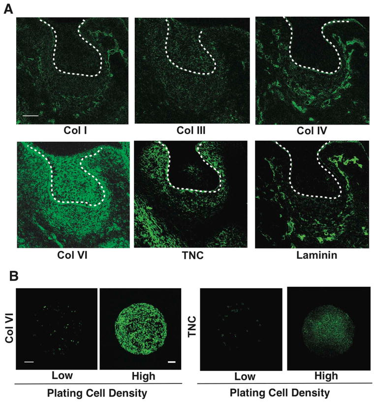
Figure 3. Natural ECM scaffold of collagen VI in condensing mesenchyme.
(A) Fluorescence micrographs showing protein levels of collagen (Col) I, III, IV, VI, tenascin C (TNC) and laminin in the tooth germ at E14. Dashed lines indicate the epithelial-mesenchymal interface, and tip of white arrows abut on the lower edge of the condensed mesenchyme. (B) Fluorescence micrographs showing protein levels of Col VI and TNC in mesenchymal cells cultured for 16 hr on FN islands (500 μm diameter) in vitro at low or high plating density (0.2 or 2.5X10⁵ cells/cm2, respectively). Scale bars represent 50 μm for A and 100 μm for B.

