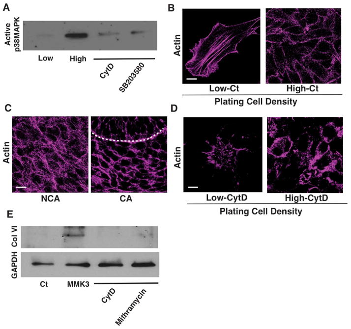
Figure 6. Synthesis of collagen VI is mediated by the mechanosensitive actin-p38MAPK-SP1 signaling pathway.
(A) Immunoblots showing active p38MAPK in mesenchymal cells cultured at low or high plating density with or without CytD (2.5 μM) or SB20358 (500 nM). (B) Fluorescence micrographs showing actin filaments in the mesenchymal cells cultured at low and high plating density in vitro. (C) Fluorescence micrographs showing actin filaments in the mesenchymal cells of non-condensed area (NCA) and condensed area (CA) in E14 tooth germ, respectively. Dashed lines indicate the epithelial-mesenchymal interface. (D) Fluorescence micrographs showing actin filaments in the mesenchymal cells cultured at low and high plating density with or without CytD (2.5 μM) in vitro. (E) Immunoblots showing collagen (Col) VI and GAPDH levels in the mesenchymal cells (Ct or MMK3 transfected), treated with CytD (2.5 μM) or mithramycin (250 nM). Scale bars represent 20 μm for C and 10 μm for B and D.

