
Figure 2.
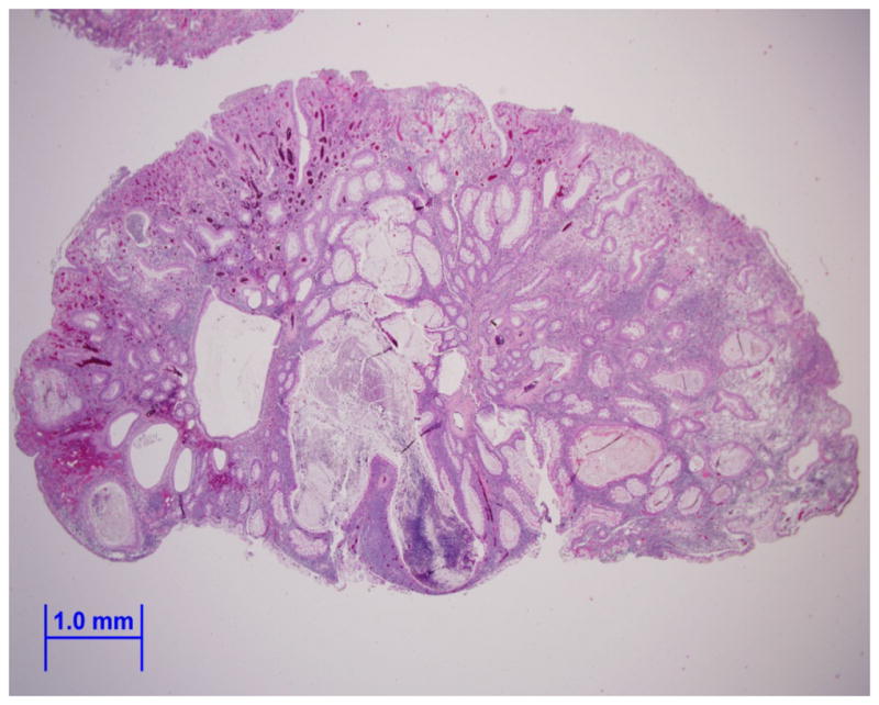

Image (A) is an endoscopically removed juvenile polyp from our patient showing a smooth surface with dilated mucus-filled cystic glands in the lamina propria (hematoxylin-eosin, x10). Image (B) shows a view of the dense stroma, inflammatory infiltrate, and fibrovascular core of the juvenile polyp (hematoxylin-eosin, x20).