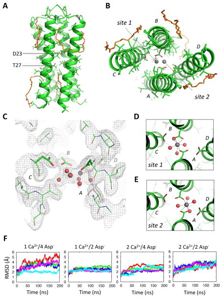
Figure 2. Crystal structure of a DAP12-TM tetramer with coordinated calcium at 2.14 Å resolution.
Side (A) and top (B) views of the tetrameric DAP12-TM crystal structure. Structured monoolein molecules are shown in orange stick representation. Two possible calcium coordination sites are indicated in (B). (C) Electron density depicted by a 2mFo-DFc map (sigma level 1.0) around the aspartic acid and threonines. (D–E) The extra density at this site was modelled as two overlapping Ca2+ coordination sites with pentagonal bipyramidal geometry, each with 50% occupancy, that included a network of additional water molecules. (F) 200-ns fully atomistic molecular dynamics simulations of the tetrameric structure with one or two Calcium ions (1Ca2+, 2Ca2+) and four or two ionized aspartic acid residues (4Asp−, 2Asp−) were run in an explicit bilayer composed of POPC molecules with 50 mM CaCl2 in the bulk solvent. Each plot shows the time-series of the RMSD deviation from the starting structure for five independent trajectories.

