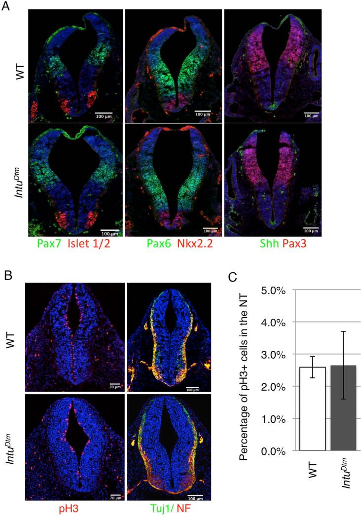
Figure 4. Neural patterning, proliferation, and differentiation are not obviously affected in IntuDtm mutant embryos.
(A) Dorsal/ventral patterning in the anterior neural tube of E10.5 IntuDtm mutant embryos is not apparently affected as shown by staining for Shh, Nkx2.2, Isl1/2, Pax6 and Pax7 as labeled. DAPI-stained nuclei are blue. N= 4 mutant and 4 control embryos. (B) Proliferation (assessed by phospho-histone H3/pH3) and differentiation (assessed by TuJ1 and Neurofilament/NF staining) appear normal in E10.5 IntuDtm mutant neural tube. N= 2 mutant and 2 control embryos. (C) The percentage of cells in the neural tube that are pH3 positive.

