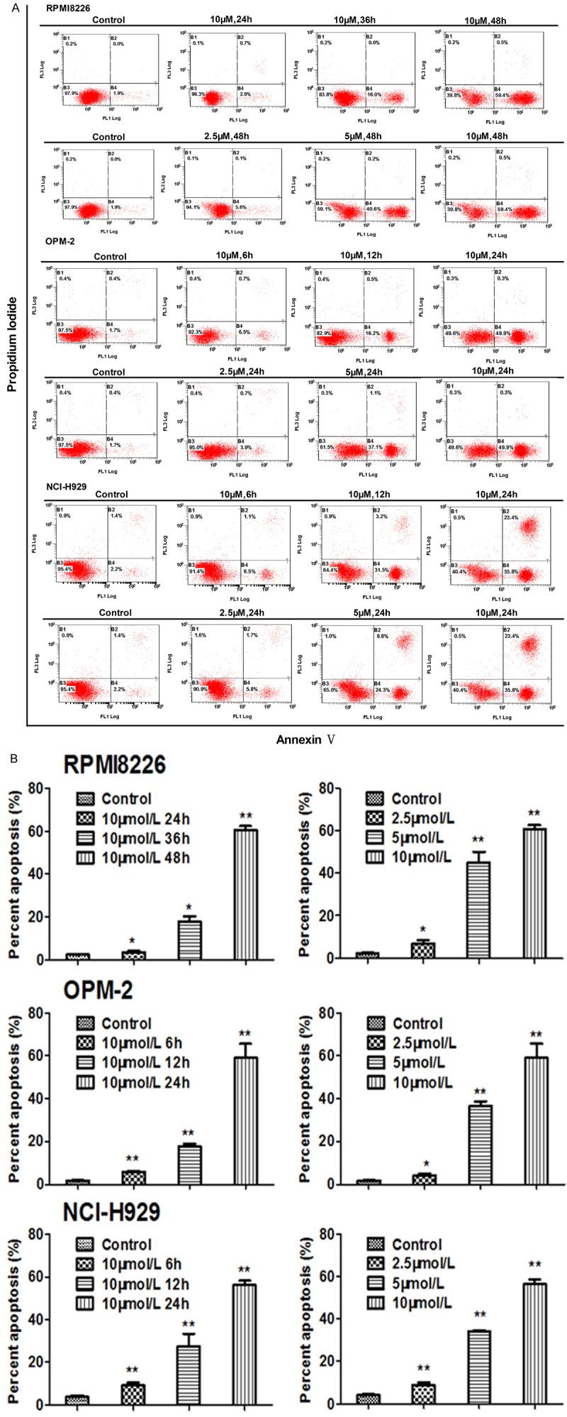
Figure 5.

HM910 induced MM cell apoptosis. A. Apoptosis analysis was performed using Annexin V/PI double staining. After cells were exposed to HM910 (2.5, 5 and 10 μmol/L), the attached and detached cells were collected. Following staining with Annexin V and PI, cells were subjected to flow cytometry analysis. B. Percentage of apoptosis. Early apoptotic cell population increased in HM910 treated myeloma cells, in a time- and dose-dependent manner. Data are presented as mean ± SD from three independent experiments (*, P < 0.05; **, P < 0.01).
