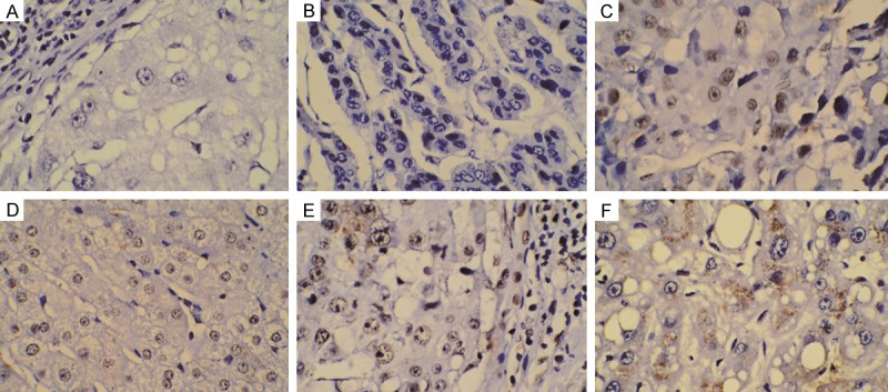
Figure 1.

Immunohistochemical staining of Musashi-2(MSI2) expression in normal tissue and hepatocellular carcinoma. (A, B) Negative MSI2 expression in normal hepatic tissue (A) and primary tumor tissue (B). (D, E) Positive MSI2 staining in normal hepatic tissue (D) and poorly differentiated tumor tissue (E). Strongly Positive MSI-2 staining in nucleus (C) and cytoplasm (F) of poorly differentiated tumors. Original magnification × 400.
