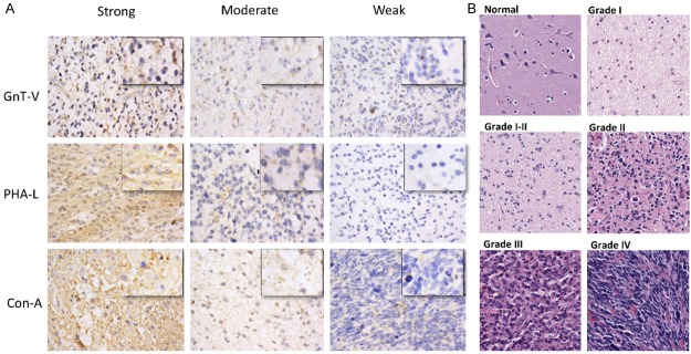
Figure 1.

Immuno-staining of different histological stages of astrocytomas. A. Representative micrographs of immunohistochemical staining of astrocytic glioma tissue samples with GnT-V antibody, PHA-L and Con-A lectins. Staining intensities are divided into three tired grading systems for each group, independently. The staining intensity of every group was calculated by scanning each core of tissue microarray by Aperio image scope and the final positivity of each sample was calculated by the formula [A + B + C / Z+ (A + B + C)], where A = Total Intensity of weak positive, B = Total Intensity of Moderate Positive, C = Total intensity of Strong Positives, Z = Total intensity of negative. Original magnification was (X400; Leica DM400B microscope). B. Representative micrographs of H&E stained tissue samples of different grades of astrocytic glioma and normal brain tissue. Original magnification was X200. Tumor grades were determined according to the 2007 WHO grading system by the providers.
