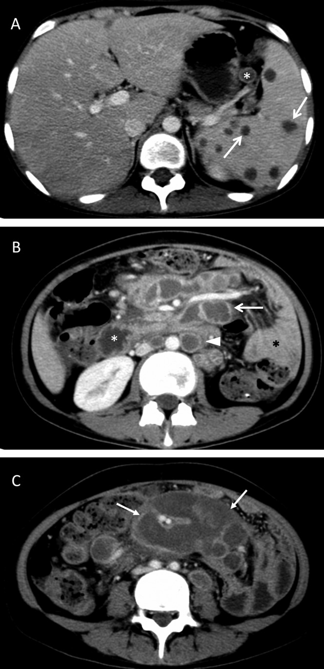
Figure 1.
A 19-year-old female with abdominopelvic and splenic tuberculosis. A: A contrast-enhanced image shows peripheral enhancement of enlarged lymph nodes in the splenic hilum (the lesser omentum; star) and sporadic peripheral enhancement lesions in the spleen (arrow). B: A contrast-enhanced image shows peripheral enhancement of enlarged lymph nodes in the anterior pararenal space (the mesenteric root; arrow) and the upper para-aortic region (arrowhead). The descending duodenum can be recognized by its mucosal folds (white star). The adjacent intestinal tract shows homogeneous density (black star). C: The enlarged lymph nodes demonstrate peripheral enhancement with a multilocular appearance in the mesentery (arrow).

