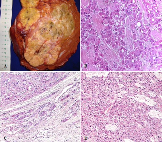
Figure 2.

Pathological aspects of fibrolamellar hepatocellular carcinoma. (A) Surgical specimen of a large, well-circumscribed, yellow, heterogeneous mass with areas of necrosis and a central scar. (B) Histopathology of “pure fibrolamellar hepatocellular carcinoma”: large tumor cells with deeply eosinophilic cytoplasm, round, central macronucleoli, and abundant fibrous stroma arranged in thin parallel lamellae around the tumor cells. (C): Fibrolamellar hepatocellular carcinoma showing microvascular invasion; (D): “Mixed fibrolamellar hepatocellular carcinoma”: classical fibrolamellar hepatocellular carcinoma cells are found intermingled with neoplastic cells with features of conventional hepatocellular carcinoma.
