Fig. 5.
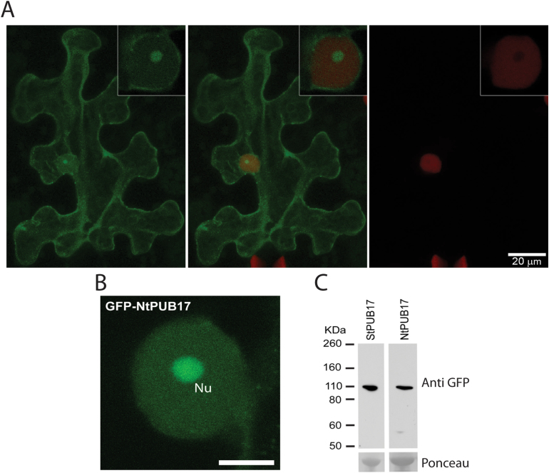
GFP-StPUB17 and GFP-NtPUB17 strongly accumulate in the nucleolus in planta. (A) Representative CB157 N. benthamiana leaves expressing mRFP-H2B examined by confocal microscopy 48h after infiltration of 35S-GFP-StPUB17, indicating green (left), red (right), and merged (centre) channels, with close-up nuclear confocal images inset. (B) Representative close-up nuclear confocal image showing the sub-nuclear localization of GFP-NtPUB17. Nu indicates the nucleolus and the scale bar is 10 μm. (C) Western blots probed with a GFP antibody showing stable protein fusions of potato and tobacco GFP-PUB17 of the expected size. The lower panels show Ponceau staining of the membrane as a loading control.

