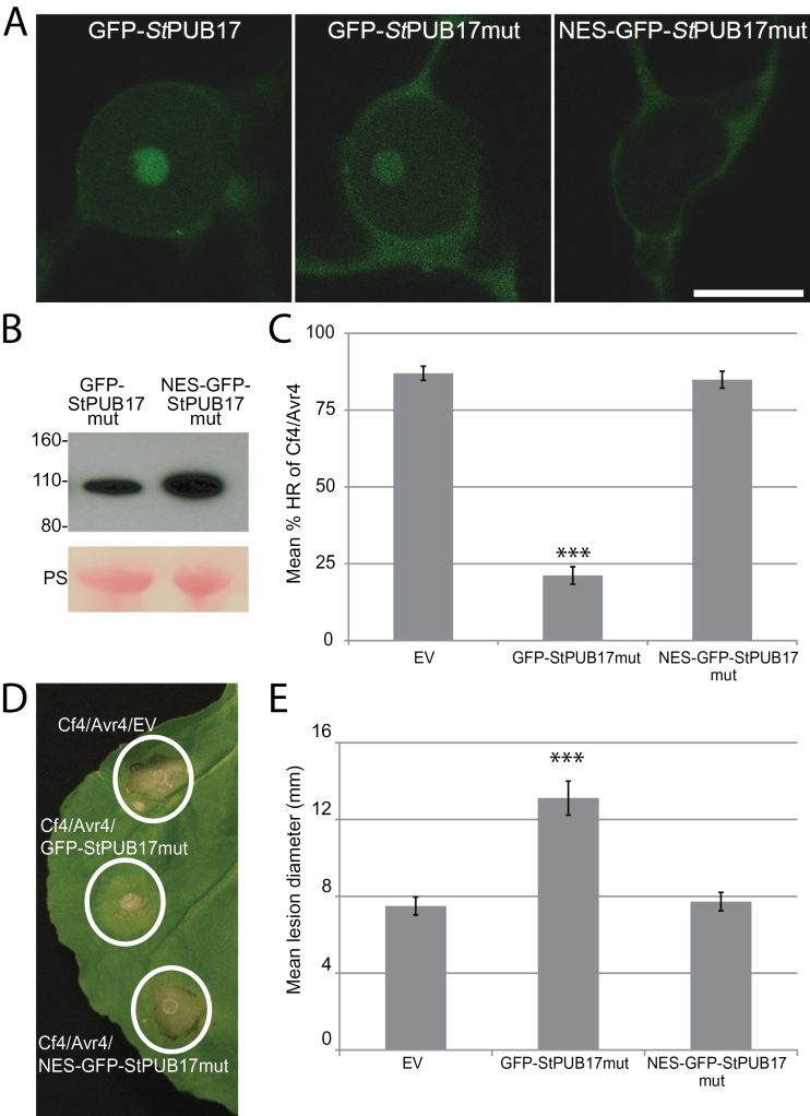
Fig. 6.
Dominant-negative activity of an StPUB17V314I,V316I mutant (StPUB17mut) is prevented by nuclear exclusion. (A) Representative nuclear images of GFP-STPUB17, GFP-StPUB17mut, and NES-GFP-StPUB17mut. Size marker is 10 μm. (B) Western blots probed with a GFP antibody showing stable protein fusions of GFP-StPUB17mut and NES-GFP-StPUB17mut of the expected size. The lower panels show Ponceau staining (PS) of the membrane as a loading control. (C) Mean % HR of Cf4/Avr4 co-expressed with empty vector (EV), GFP-StPUB17mut, and NES-GFP-StPUB17mut. (D) Images of HR of Cf4/Avr4 co-expressed with empty vector (EV), GFP-StPUB17mut, and NES-GFP-StPUB17mut. (E) Mean lesion diameter of P. infestans colonization following expression of empty vector (EV), GFP-StPUB17mut, and NES-GFP-StPUB17mut. Statistical analysis was carried out using ANOVA with pairwise comparisons performed with a Holm–Sidak test; ***P ≤0.001 [n=92 for (C) and n=106 for (E)]; error bars show standard error.

