
Fig. 3.
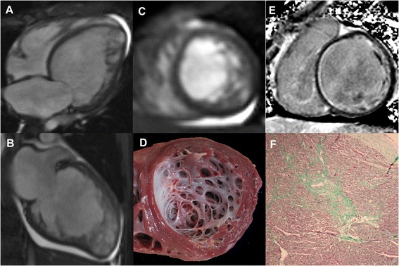
Representative images in a 14-year-old boy with LVNC underwent heart transplantation. The horizontal (a) and vertical (b) long-axis and short-axis (c) end-diastolic cine images showed prominent trabeculations in anterior, inferior, and lateral segments at apical level. The apical short-axis section of explanted heart (d) revealed prominent trabeculations. Contrast-enhanced image (e) demonstrated subendocardial LGE in the basal septum. Masson’s Trichrome staining (f) confirmed the present of replacement fibrosis in corresponding subendocardial area of basal septum (green area); magnification × 50