Fig. 4.

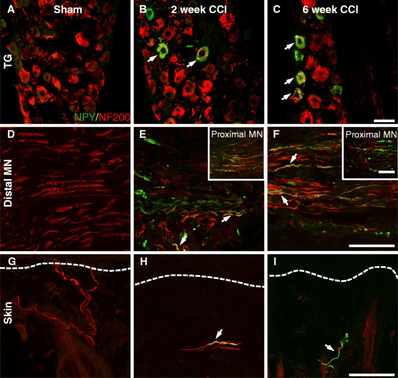
NPY was expressed by some NF200-IR sensory neurons after CCI. The presence of NPY immunoreactivity (in green) in NF200-positive sensory fibers (in red) was assessed in sham animals and in CCI animals 2 and 6 weeks after injury in the trigeminal ganglia (TG), the mental nerve (MN) and in the upper dermis of the skin. Colocalization between these two markers is shown in yellow and highlighted with an arrow. NPY was not seen in the TG of sham animals (a), but was expressed in some NF200-IR cell bodies 2 (b) and 6 weeks (c) after CCI. The percentage of NF200-IR cells that expressed NPY was quantified (Sham 0 ± 0; 2 week CCI 28.7 ± 5.4 %; 6 week CCI 21.1 ± 2.5 %; n = 6). The percentage of NPY-IR cells that expressed NF200 was quantified (Sham 0 ± 0; 2 week CCI 57.5 ± 3.8 %; 6 week CCI 48.5 ± 5.1 %; n = 6). No NPY-IR fibers were seen in the MN of sham animals (d). Two (e) and 6 weeks (f) after CCI some NF200 fibers distal to the ligation expressed NPY. NPY was also seen proximal to the ligation (inset in e-f). In the skin, NF200 fibers were present in the upper dermis of sham animals (g). NF200 fiber density was reduced in the skin after CCI and some NF200 fibers expressed NPY immunoreactivity 2 (h) and 6 weeks (i) after CCI. Dashed lines represent the border between the epidermis and the upper dermis. In all cases, 6 week sham animals were used as representative images and data from 2 and 6 week sham animals were pooled for quantification. Scale bar = 50 μm TG: trigeminal ganglia, MN: mental nerve
